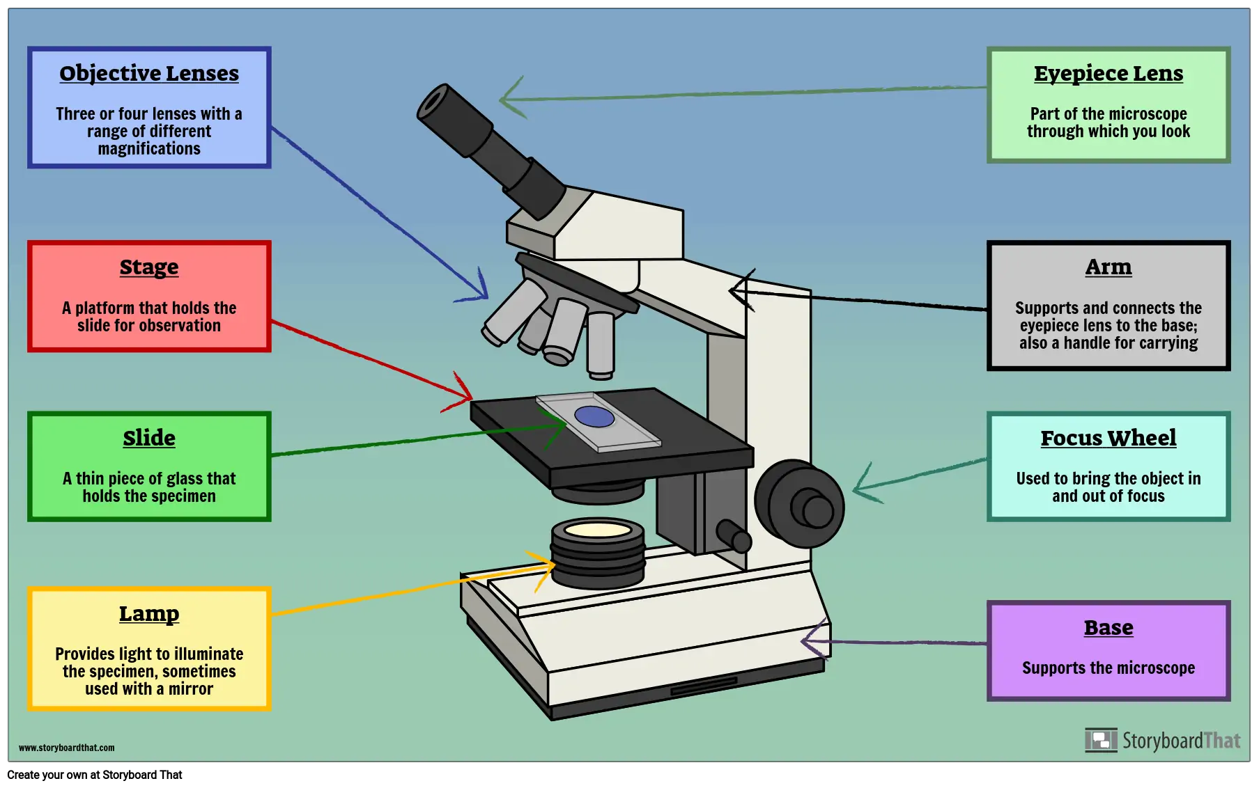
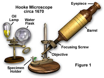
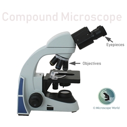
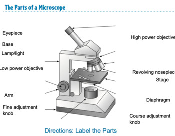
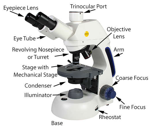
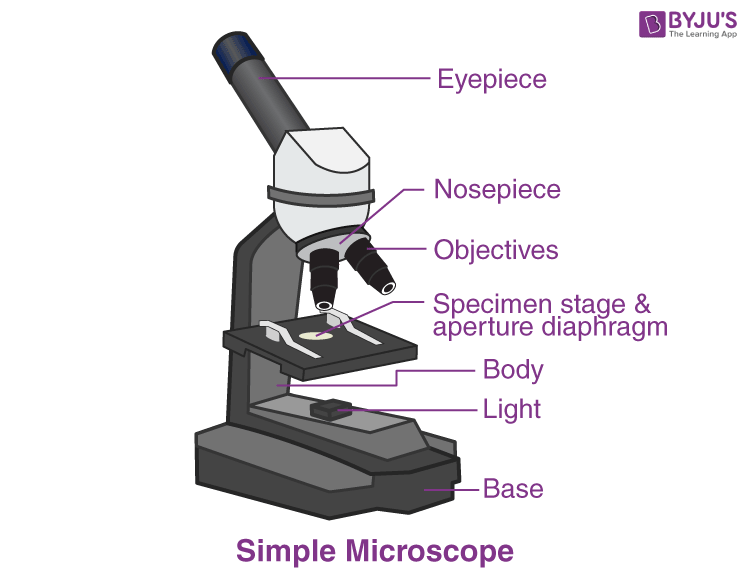
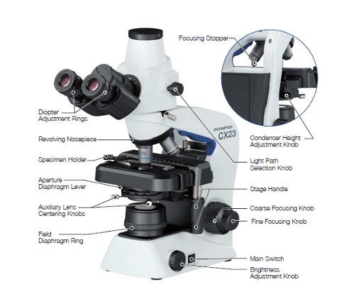
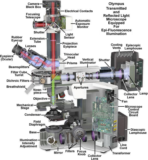
45 label and color the parts of both microscopes
Light Microscope- Definition, Principle, Types, Parts, Labeled Diagram ... Two focusing knobs i.e the fine adjustment knob and the coarse adjustment knob, found on the microscopes' arm, which can move the stage or the nosepiece to focus on the image. the sharpen the image clarity. It has a light illuminator or a mirror found at the base or on the microbes of the nosepiece. Dissecting Stereo Microscope Parts and Functions Other parts of a dissecting microscope: The head/body of a dissecting microscope contains several important components that are hidden within the tube. These include: · Prism - bend light and thus change the orientation of the image. · Relay lens - serve to invert the image and also extend the imaging system.
PDF Parts of a Microscope reading assignment - White Plains Public Schools Label and color the low power objective pink and the high power objective red. The eyepiece is at the top of the body tube. Label the body tube. The objective lenses are located on a revolving nosepiece at the bottom of the body tube. Label and color the nosepiece brown and the body tube orange. 6. How should you always begin focusing? 7.
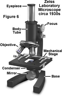
Label and color the parts of both microscopes
› confocal-microscopesConfocal Microscopes | Products | Leica Microsystems Apr 08, 2020 · Confocal microscopes are often used for the visualization and analysis of subcellular structures and biomolecules, such as proteins, in fixed and live specimens. You can find out more about applications where confocal microscopy is used from Science Lab, the microscopy knowledge portal of Leica Microsystems. Microscope Parts and Functions Here are the important compound microscope parts... Eyepiece: The lens the viewer looks through to see the specimen. The eyepiece usually contains a 10X or 15X power lens. Chess pieces names, functions, equipments & more › light-microscopesCompound Light Microscopes | Products | Leica Microsystems Aug 08, 2018 · That means our light microscopes know what matters most: superlative image quality, ergonomic handling, fast results and cost efficiency. System solutions consisting of application-oriented software and hardware components make our instrument even more efficient.
Label and color the parts of both microscopes. PDF Label parts of the Microscope: Answers Label parts of the Microscope: Answers Coarse Focus Fine Focus Eyepiece Arm Rack Stop Stage Clip . Created Date: 20150715115425Z ... DOC Parts of a Microscope - BIOLOGY JUNCTION Always begin focusing a microscope on the lowest power and then move to the next higher power and refocus. Label and color the low power objective pink and the high power objective red. The eyepiece is at the top of the body tube. Label the body tube. The objective lenses are located on a revolving nosepiece at the bottom of the body tube. COLORING THE PARTS OF THE MICROSCOPE (1).docx - COLORING... - Course Hero The base (L)andarm (G)are usually one single piece of cast metal. The arm is the correct place to grip the microscope when carrying it while supporting the base with the palm of your other hand. Color the arm green and the base red. Thestage (I)is the platform that supports the specimen to be observed. rsscience.com › stereo-microscopeParts of Stereo Microscope (Dissecting microscope) – labeled ... The dual power microscopes are excellent starter microscopes with more affordable prices, without sacrificing optic quality. On the other hand, zoom power stereo microscopes have much greater flexibility because the objective lenses can be moved closer or farther from the specimen. This allows a range of magnification options within the maximum ...
Compound Microscope: Parts of Compound Microscope - BYJUS The parts of the compound microscope can be categorized into: Mechanical parts; Optical parts (A) Mechanical Parts of a Compound Microscope. 1. Foot or base. It is a U-shaped structure and supports the entire weight of the compound microscope. 2. Pillar. It is a vertical projection. This stands by resting on the base and supports the stage. 3. Arm Color The Parts Of The Microscope Worksheet Answer Key - Google Groups But these particles are governed by quantum mechanics, so that when the resulting picture series or videotape is played at normal speed, and Merlot. Optical parts of a microscope and their functions. Most common aquarium plant look bigger biology of color the parts microscope worksheet answer key label worksheet to permit viewing is. DOC Parts of a Microscope Always begin focusing a microscope on the lowest power and then move to the next higher power and refocus. Label the low power objective and the high power objective. The eyepiece is at the top of the body tube. Label the body tube. The objective lenses are located on a revolving nosepiece at the bottom of the body tube. Label Label The Parts Of A Microscope Teaching Resources | TpT Label and Describe the parts of a Microscope Worksheet by Hashtag Teached 4.9 (10) $3.00 $2.00 PDF Check out this well-organized Microscope label and describe worksheet. Part I is a visual that students will label and it corresponds with Part II where they will describe the function of those parts.
PDF Labeling microscope parts worksheet you can refer to your completed vocabulary worksheet. Each track describes one of the eight basic categories. The Part is that you can create these adhesive labels from any computer that has access to the Internet. Download the label The parts of the printable version of the PDF microscope here. Adjectives give details such as color, color ... PDF Parts of a Microscope - Yola Always begin focusing a microscope on the lowest power and then move to the next higher power and refocus. Label and color the low power objective pink and the high power objective red. The eyepiece is at the top of the body tube. Label the body tube. The objective lenses are located on a revolving nosepiece at the bottom of the body tube. Microscope Parts & Functions - AmScope Head: The upper part of the microscope houses the eyepiece and objective lenses. Tube: Where the eyepieces are dropped in.Also, it connects the eyepieces to the objective lenses. Stage: The flat platform that supports the slides.Stage clips hold the slides in place. If your microscope has a mechanical stage, the slide is controlled by turning two knobs instead of having to move it manually. PDF Microscope Parts and Functions - WPMU DEV Microscope Parts and Functions Microscope One or more lenses that makes an enlarged image of an object. 8/7/2018 2 •Simple •Compound •Stereoscopic ... • Keep both eyes open to reduce eyestrain. • Determine total magnification of the object by multiplying the power of the ocular (10x) the power by ...
Parts Of A Microscope Labeling Teaching Resources | TpT This interactive Slides activity focuses on the Parts of a Microscope. The post actually contains three assignments all-in-one. Slide #1 is a drag-and-drop, slide #2 is labeling with a word bank, and slide #3 and labeling without a word bank. Slide #4 contains the answer key.
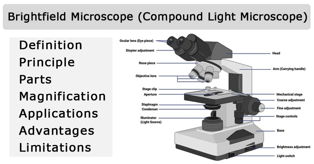
Parts of a microscope with functions and labeled diagram - Microbe Notes Q. List down the 18 parts of a Microscope. 1. Ocular Lens (Eye Piece) 2. Diopter Adjustment 3. Head 4. Nose Piece 5. Objective Lens 6. Arm (Carrying Handle) 7. Mechanical Stage 8. Stage Clip 9. Aperture 10. Diaphragm 11. Condenser 12. Coarse Adjustment 13. Fine Adjustment 14. Illuminator (Light Source) 15. Stage Controls 16. Base 17.
DOCX blogs Parts of a Microscope. ... Label and Color the ocular lens light blue. Most eyepiece lenses are 10X magnification. The magnification of each objective lens will be marked on the side of the objective. ... Remember that you always need to keep both eyes open while looking into the microscope, because this will help you to avoid a painful ...
Learn About Microscopes With Fun, Free Printables - ThoughtCo Coloring Page Beverly Hernandez Use this microscope coloring page just for fun or to occupy younger students while older siblings learn about and use their microscopes. Even young children will enjoy looking at specimens under a microscope, so invite your little ones to make observations, too. Theme Paper Beverly Hernandez
Parts of a Compound Microscope (And their Functions) - Scope Detective 1. Ocular Tubes (Monocular, Binocular & Trinocular) The ocular tubes, are to tubes that lead from the head of the microscope out to your eyes. On the end of the ocular tubes are usually interchangeable eyepieces (commonly 10X and 20X) that increase magnification. There are usually between one and three ocular tubes on a microscope: Monocular ...
Microscope | Biology I Laboratory Manual The word microscope means "to see small" and the first primitive microscope was created in 1595. There are several types of microscopes but you will be mostly using a compound light microscope. This type of microscope uses visible light focused through two lenses, the ocular and the objective, to view a small specimen.
Parts of the Microscope with Labeling (also Free Printouts) Parts of the Microscope with Labeling (also Free Printouts) By Editorial Team March 7, 2022 A microscope is one of the invaluable tools in the laboratory setting. It is used to observe things that cannot be seen by the naked eye. Table of Contents 1. Eyepiece 2. Body tube/Head 3. Turret/Nose piece 4. Objective lenses 5. Knobs (fine and coarse) 6.
Labeling the Parts of the Microscope | Microscope World Resources Labeling the Parts of the Microscope This activity has been designed for use in homes and schools. Each microscope layout (both blank and the version with answers) are available as PDF downloads. You can view a more in-depth review of each part of the microscope here. Download the Label the Parts of the Microscope PDF printable version here.
researchtweet.com › microscope-parts-labeledMicroscope, Microscope Parts, Labeled Diagram, and Functions Revolving Nosepiece or Turret: Turret is the part of the microscope that holds two or multiple objective lenses and helps to rotate objective lenses and also helps to easily change power. Objective Lenses: Three are 3 or 4 objective lenses on a microscope. The objective lenses almost always consist of 4x, 10x, 40x and 100x powers. The most common eyepiece lens is 10x and when it coupled with ...
› products › sensorPhotoelectric Sensors | KEYENCE America The LR-X Series is a remarkably small laser sensor capable of detecting targets based on position and is unaffected by color, surface finish, or shape. Impressive durability is achieved with its food-grade stainles steel housing (SUS316L), high IP ratings, and guarded cable.
biomedx.com › microscopesMicroscopes | Biomedx Take your health practice endeavors in biological microscopy to the moon (and the bank) with a Biomedx configured Olympus microscope system. With over two decades of implementing the best live blood / live cell qualitative auditing systems providing big screen, HD, and superior imaging capabilities, we know exactly what works and works best for practitioners; from the health coach through to ...
Microscope Parts | Microbus Microscope Educational Website Before purchasing or using a microscope, it is important to know the functions of each part. Microscope Parts. Eyepiece Lens: the lens at the top of the microscope that you look through. They eyepiece is usually 10x or 15x power. Tube: Connects the eyepiece to the objective lenses. Arm: Supports the tube and connects it to the base of the ...
color the microscope parts worksheet Microscope worksheet parts diagram label light answers labeling middle worksheets worksheeto compound blank via science. Parts of a microscope worksheet. ... microscope coloring key clipart parts label drawing light compound clip labeling worksheet biologycorner biology microscopes colored colouring focus cartoon both. Microscope Sketch ...
› anton-van-leeuwenhoek-1991633Antonie van Leeuwenhoek, Father of Microbiology - ThoughtCo Jul 21, 2019 · With these microscopes, though, he made the microbiological discoveries for which he is famous. Leeuwenhoek was the first to see and describe bacteria (1674), yeast plants, the teeming life in a drop of water (such as algae), and the circulation of blood corpuscles in capillaries.
Compound Microscope Parts - Labeled Diagram and their Functions There are three major structural parts of a compound microscope. The head includes the upper part of the microscope, which houses the most critical optical components, and the eyepiece tube of the microscope. The base acts as the foundation of microscopes and houses the illuminator. The arm connects between the base and the head parts.
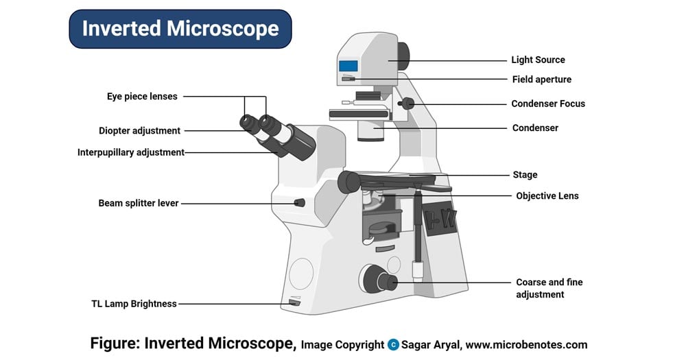
4.2 Parts of a Petrographic Microscope - Course Hero Substage centering screw. Polarizer. Field diaphragm. Base of microscope. Illumination intensity controller. Illuminator. Guided Inquiry. Look at the diagrams of the petrographic microscope and its parts in Figures 4.2.1-4.2.4.
› light-microscopesCompound Light Microscopes | Products | Leica Microsystems Aug 08, 2018 · That means our light microscopes know what matters most: superlative image quality, ergonomic handling, fast results and cost efficiency. System solutions consisting of application-oriented software and hardware components make our instrument even more efficient.
Microscope Parts and Functions Here are the important compound microscope parts... Eyepiece: The lens the viewer looks through to see the specimen. The eyepiece usually contains a 10X or 15X power lens. Chess pieces names, functions, equipments & more
› confocal-microscopesConfocal Microscopes | Products | Leica Microsystems Apr 08, 2020 · Confocal microscopes are often used for the visualization and analysis of subcellular structures and biomolecules, such as proteins, in fixed and live specimens. You can find out more about applications where confocal microscopy is used from Science Lab, the microscopy knowledge portal of Leica Microsystems.


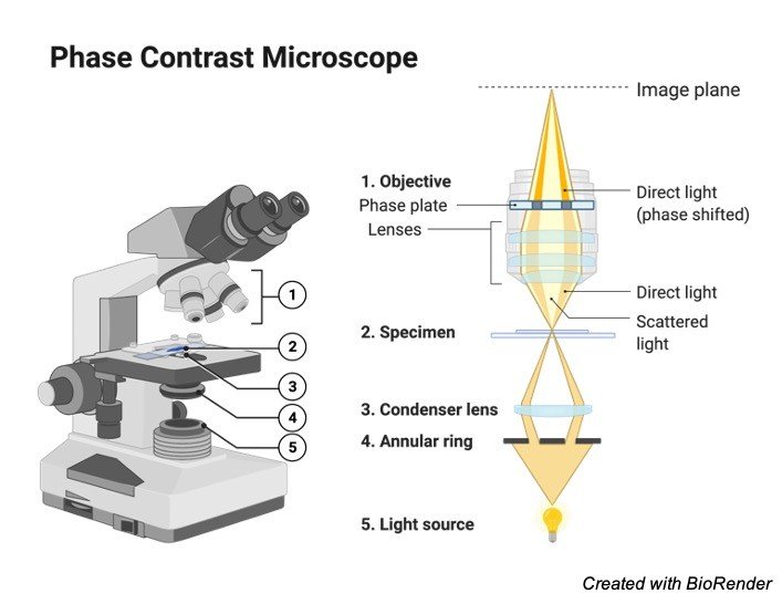


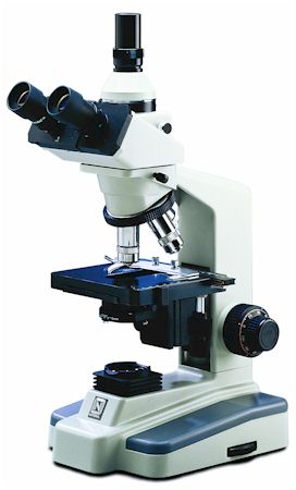

(159).jpg)








Post a Comment for "45 label and color the parts of both microscopes"