43 neuron model with labels
A Guide to Understand Neuron with Neuron Diagram | EdrawMax Online To properly understand the coordination between the brain and the body, the students must learn about the neurons. They can use neuron-labeled diagrams while learning the complex structure of neurons. Creating a neuron-labeled image by hand can be difficult. The students must use the EdrawMax Online tool to make a high-quality neuron diagram . 2. Label a Neuron Model Quiz - purposegames.com This is an online quiz called Label a Neuron Model There is a printable worksheet available for download here so you can take the quiz with pen and paper. From the quiz author Practice labeling structures of a neuron model. Your Skills & Rank Total Points 0 Get started! Today's Rank -- 0 Today 's Points One of us! Game Points 11
Neuron Model Labeling Diagram | Quizlet Neuron Model Labeling. STUDY. Learn. Flashcards. Write. Spell. Test. PLAY. Match. Gravity. Created by. lindsey_lasater0 PLUS. Terms in this set (7) ... is the spherical part of the neuron that contains the nucleus. The cell body connects to the dendrites, which bring information to the neuron, and the axon, which sends information to other ...
Neuron model with labels
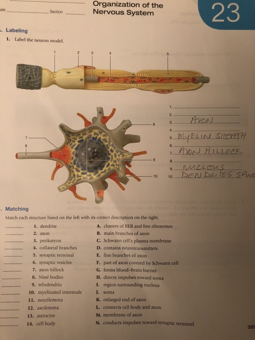
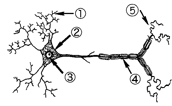
Nervous system anatomy, Medical anatomy, Neuron model Mar 12, 2018 - classroom.sdmesa.edu anatomy images Nervous_label neuron2_label_label.jpg Solved Label the neuron model | Chegg.com Label the neuron model; Question: Label the neuron model. This problem has been solved! See the answer See the answer See the answer done loading. Label the neuron model. Expert Answer. Who are the experts? Experts are tested by Chegg as specialists in their subject area. We review their content and use your feedback to keep the quality high. Diagram Quiz on Neuron Structure and Function (Labeling Quiz) 1. Identify the cell type in the above figure Liver Cell Cardiac Cell Nerve cell Skin cell 2. In the figure, labeled '1' receives impulses from adjacent neuron. It is called the Dendron Dendrite Axon Axonite 3. In the figure, labeled '2' is the short filaments from the cell body that carries impulses from dendrites to the cell body which is the
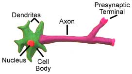
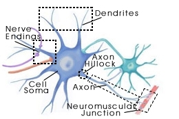
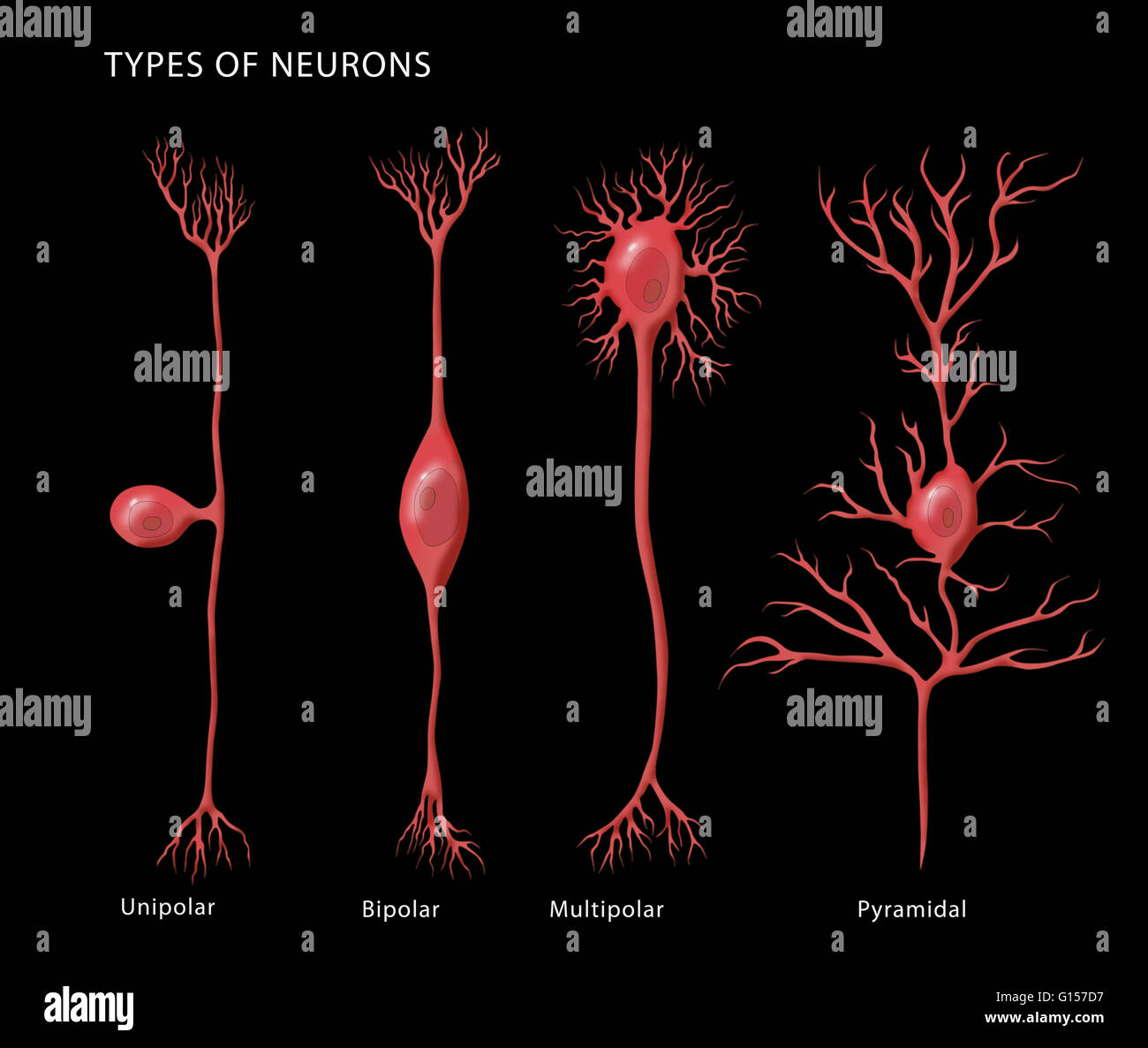
Neuron model with labels. Neuron (Nerve Cell) Types, Structure and Function A relay neuron (also known as an interneuron) allows sensory and motor neurons to communicate with each other. Relay neurons connect various neurons within the brain and spinal cord, and are easy to recognize, due to their short axons. Alike to motor neurons, interneurons are multipolar. This means they have one axon and several dendrites. Neuron Labeling - The Biology Corner Students in anatomy and physiology spent this year learning remotely, which required some adjustments in how material was presented. In the past, students would label a nerve cell and color neuroglia cells using paper handouts to learn the structures of the neuron and the neuroglia (supporting cells). Neuroscience for Kids - Models - University of Washington Create a model of a neuron by using clay, playdough, styrofoam, recyclables, food or anything else you can get your hands on. Use pictures from books to give you an idea of where the components of a neuron should go and what shape they should be. Use different colors to indicate different structures. Make a neural circuit with a few of the Label Neuron Anatomy Printout - EnchantedLearning.com Read the definitions, then label the neuron diagram below. axon - the long extension of a neuron that carries nerve impulses away from the body of the cell. cell body - the cell body of the neuron; it contains the nucleus (also called the soma) dendrites - the branching structure of a neuron that receives messages (attached to the cell body)
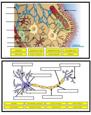
Nervous System - Label the Neuron Choose the correct names for the parts of the neuron. (1) (2) (3) (4) (5) (6) This neuron part receives messages from other neurons. (7) This neuron part sends on messages to other neurons. (8) This neuron part gives messages to muscle tissue. (9) This neuron part processes incoming messages. Biological neuron model - Wikipedia where V m is the voltage across the cell membrane and R m is the membrane resistance. (The non-leaky integrate-and-fire model is retrieved in the limit R m to infinity, i.e. if the membrane is a perfect insulator). The model equation is valid for arbitrary time-dependent input until a threshold V th is reached; thereafter the membrane potential is reset.. For constant input, the minimum input ... Neuron Models - San Diego Mesa College Neuron Models. Click on a photo for a larger view of the model. Click on Label for the labeled model. Back to Nervous System. Neuron: Cell Body & Dendrites: Axon: Label: Label: Label: Neuron: Cell Body & Dendrites: Axon: Label: Label: Neuron Model - MCCC Spinal Cord Spinal Cord Slide Neuron Model Neuron Slide. Brain Superior View Inferior View Sagittal View
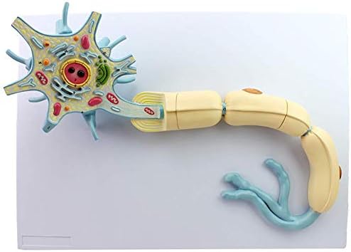
Neuron Model - Human Body Help Neuron Model Quiz yourself with the picture below. Scroll down for the answer key. There is a video at the bottom if you'd like to watch it first. Key: Nuclear envelope Nissl Bodies Axon terminal Axon hillock Axon Dendrites Axon Nucleus of a Schwann cell Myelin Sheath (Schwann Cell wrapping around Axon many times) Mitochondria Connective tissue Neuron Diagram & Types | Ask A Biologist Types of Neurons. There are many types of neurons in your body. Each type is specialized to be good at doing different things. Multipolar neurons have one axon and many dendritic branches. These carry signals from the central nervous system to other parts of your body such as your muscles and glands. Unipolar neurons are also known as sensory ... Labeled neuron cell body | Maquetas cerebro, Neuronas, Proyectos escolares Cell Model. Magnified more than 2500 times and fully three-dimensional, a neuron model is depicted in its natural setting. With the membranous envelope cut away, the cytological ultrastructure, organelles and inclusions within the cell body are depicted in contrasting colors. A section of the axon lifts off to expose the enveloping myelin ... Neuron Model - YouTube For pictures of this model with answer keys to help you study, visit: ...
Neuron 3D models - Sketchfab Neuron 3D models ready to view, buy, and download for free. Popular Neuron 3D models View all NeuroCap 32 0 4 【Neuron】Spinous Pyramidyal Cell 386 0 6 3D model Bacterium 3 31 0 1 Multipolar neuron 150 0 2 Neuron And Oligo-Dendrocytes (3d printing ready) 331 0 7 【Neuron】Spinous Stellate Cell 255 0 4 BRAIN2 1.8k 0 14 Pyramidal Neurons 91 0 1 Neuron
Neuron Model Worksheets & Teaching Resources | Teachers Pay Teachers Model of a Neuron Nervous System Activity Human Body Systems, Middle School by Dr Dave's Science $2.50 PDF A nervous system activity where students make models of a typical neuron. Labeled structures include dendrites, axons, cell body, and myelin sheath. The resource is editable so you can add other structures if desired.
Labeled Neuron Diagram | Science Trends Motor neurons are part of the central nervous system (CNS) and communicate signals from the spinal cord to the parts of the body to control their motion. For example, motor neurons send signals to the muscles in your arms causing them to contract. Motor neurons send electrical signals to your intestines so they move and churn food.
neuron model labeled Diagram | Quizlet neuron model labeled STUDY Learn Write Test PLAY Match Created by elizabethdeposada PLUS Terms in this set (11) telodendria ... nucleus of cell body ... node of ranvier ... myelin sheath ... nucleus of schwann cell ... neurilemma ... endoneurium ... axons ... axon hillock ... axon terminals ... dendrites ... Sets found in the same folder
Neuron Labelling Teaching Resources | Teachers Pay Teachers You can use this resource to label parts of a neuron as a model, diagram, or notes in your nervous system unit to instruct, explain, and facilitate student learning about neurons' design in the human body. Science from the South Doodle Docs can be used as a part of many different activities for your students, such as notes, part of an interactive n
Motor Neuron: Function, Types, and Structure | Simply Psychology There are two types of motor neurons: Lower motor neurons - these are neurons which travel from the spinal cord to the muscles of the body. Upper motor neurons - these are neurons which travel between the brain and the spinal cord. The structure of a motor neuron can be categorized into three components: the soma, the axon, and the dendrites.
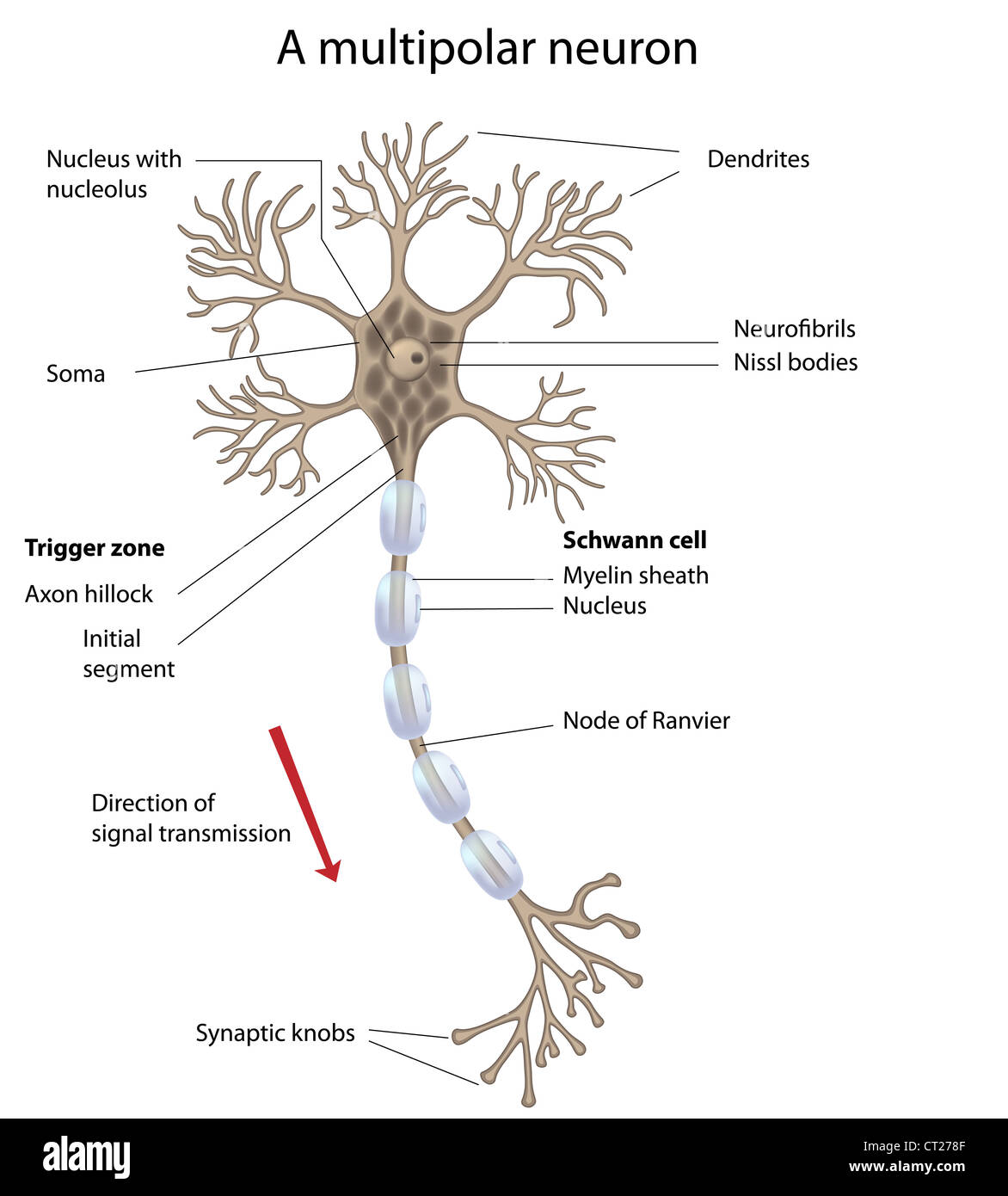
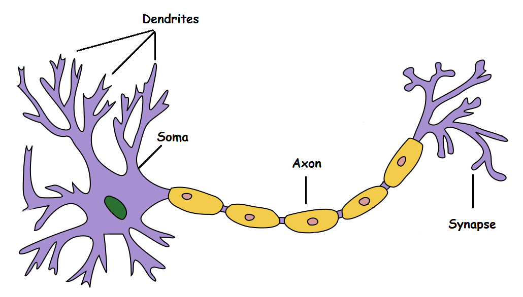
What Is a Neuron? - Definition, Structure, Parts and Function A neuron varies in shape and size depending on its function and location. All neurons have three different parts - dendrites, cell body and axon. Parts of Neuron. Following are the different parts of a neuron: Dendrites. These are branch-like structures that receive messages from other neurons and allow the transmission of messages to the ...
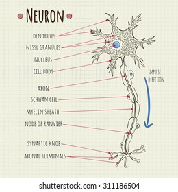
A Labelled Diagram Of Neuron with Detailed Explanations A Labelled Diagram Of Neuron with Detailed Explanations Biology Biology Article Diagram Of Neuron Diagram Of Neuron A neuron is a specialized cell, primarily involved in transmitting information through electrical and chemical signals. They are found in the brain, spinal cord and the peripheral nerves. A neuron is also known as the nerve cell.
Anatomical Models - Anatomy Teaching Models - Neuron Models - 3B Scientific Motor End Plate. $ 199.00. Item: 1000236 [C40/4] The motor end plate model depicts the neuromuscular junction with striated muscle fiber. The motor end plate of the human nervous system is depicted in colorful anatomical detail. The motor end plate model is a great addition to any lesson ... more. Add to cart.
Graph — NEURON documentation Syntax: g = new Graph () g = new Graph (0) Description: An instance of the Graph class manages a window on which x-y plots can be drawn by calling various member functions. The first form immediately maps the window to the screen. With a 0 argument the window is not mapped but can be sized and placed with the view () function.
LABELING NEURON MODEL Quiz - PurposeGames.com Playlists (1) Tournaments (37) AI Stream The more you play, the more accurate suggestions for you. Cities by Landmarks 11p Image Quiz. Cities of Midwestern US 32p Image Quiz. I spy on... 26p Image Quiz. The Western States 11p Image Quiz.
Diagram Quiz on Neuron Structure and Function (Labeling Quiz) 1. Identify the cell type in the above figure Liver Cell Cardiac Cell Nerve cell Skin cell 2. In the figure, labeled '1' receives impulses from adjacent neuron. It is called the Dendron Dendrite Axon Axonite 3. In the figure, labeled '2' is the short filaments from the cell body that carries impulses from dendrites to the cell body which is the
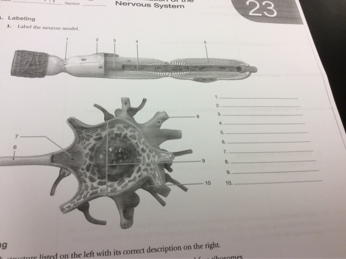
Solved Label the neuron model | Chegg.com Label the neuron model; Question: Label the neuron model. This problem has been solved! See the answer See the answer See the answer done loading. Label the neuron model. Expert Answer. Who are the experts? Experts are tested by Chegg as specialists in their subject area. We review their content and use your feedback to keep the quality high.
Nervous system anatomy, Medical anatomy, Neuron model Mar 12, 2018 - classroom.sdmesa.edu anatomy images Nervous_label neuron2_label_label.jpg







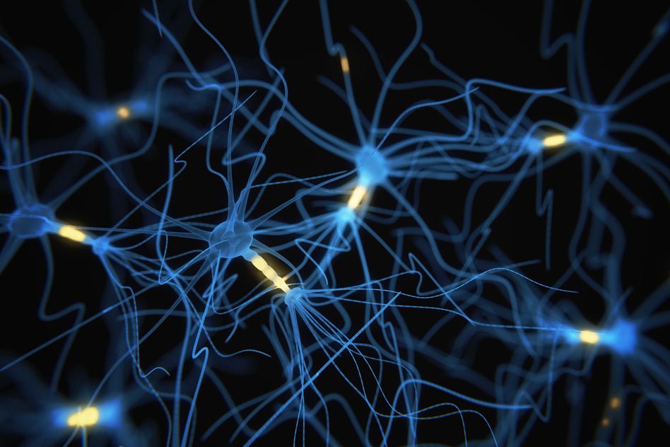


/548299803-56a796543df78cf7729765c8.jpg)



Post a Comment for "43 neuron model with labels"