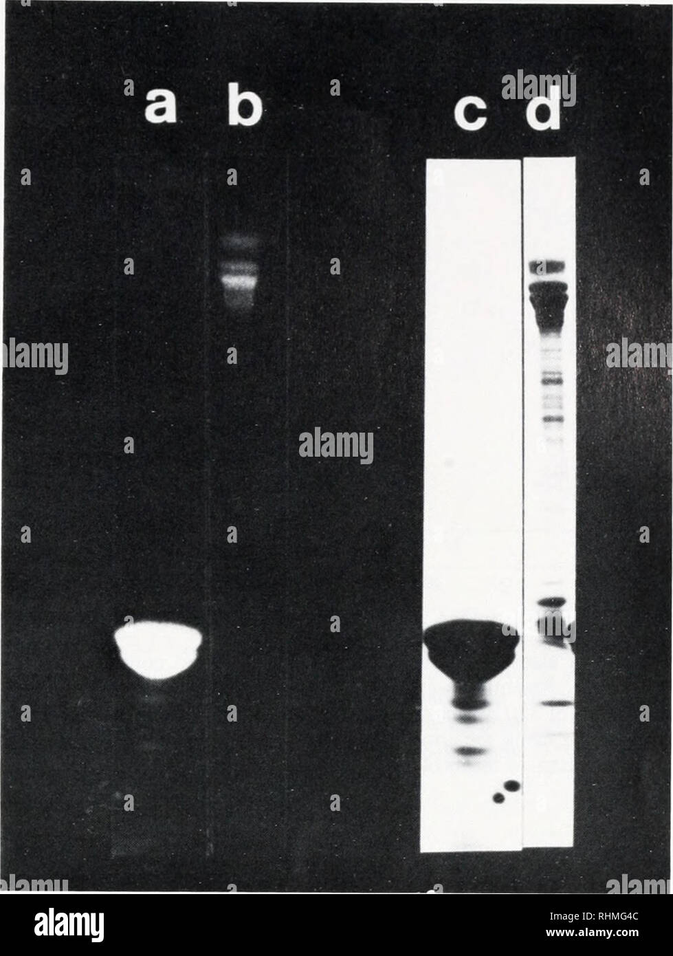
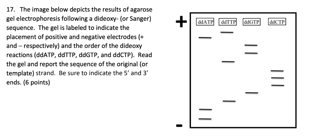
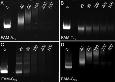
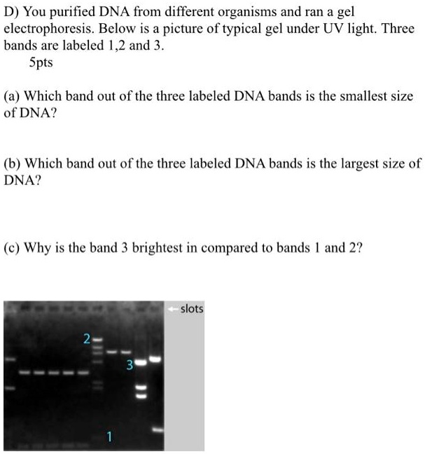
45 labeled gel electrophoresis photo
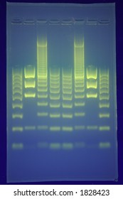
Part 2: Analyzing and Interpreting (Agarose) Gel Electrophoresis Results The gel image above is the result of restriction digestion. Lane 3, 5, 7, and 8 are a homozygous normal allele with a 184bp band here one band of 68bp is also present, but it is not visible. Lane 2 is a mutant uncut allele of 252bp. Lane 1 and 6 are heterozygous contain three alleles: 252bp, 184bp and 68bp. gel electrophoresis | Britannica The gel electrophoresis apparatus consists of a gel, which is often made from agar or polyacrylamide, and an electrophoretic chamber (typically a hard plastic box or tank) with a cathode (negative terminal) at one end and an anode (positive terminal) at the opposite end. The gel, which contains a series of wells at the cathode end, is placed inside the chamber and covered with a buffer solution.
PDF Lab 4: Gel Electrophoresis - Vanderbilt University Gel electrophoresis is a method of separating DNA fragments by movement through a Jello-like substance called agarose. Derived from a seaweed polysaccharide, agarose gels form small pores ... terms are labeled on the gel, and the loading key is labeled according to each lane. 1000bp 500bp 2000bp 250bp 100bp Lane 4 Lane Sample 1 DNA Ladder
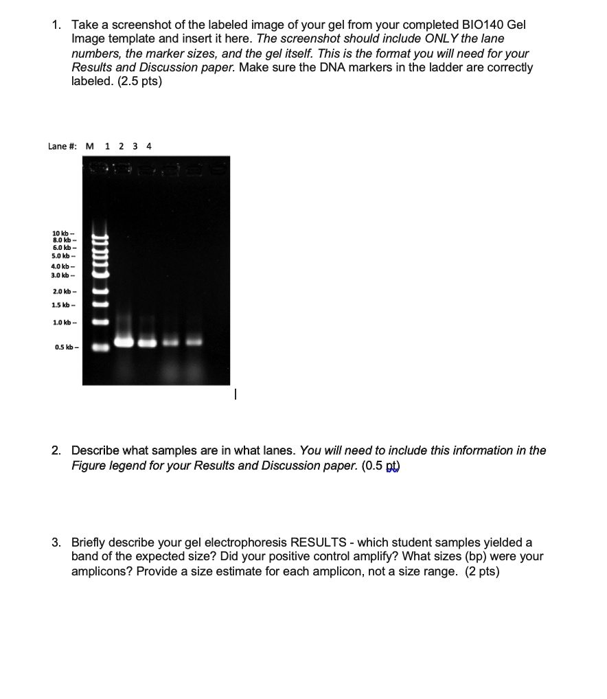
Labeled gel electrophoresis photo
Gel Electrophoresis: Molecular Biology Science Activity | Exploratorium ... Prepare your gel: Make a 0.2% sodium bicarbonate buffer by dissolving 2 grams of baking soda in 1 liter of water. You will need approximately 100 milliliters per set up—half to make the gel and half to run your samples. Make a 1% gel solution by adding 0.5 g of agar-agar powder to 50 mL of sodium bicarbonate buffer. Annotating A Gel | Get Your Science On Wiki | Fandom 1.In Inkscape import your gel file and adjust the size of your picture to fit the page out line (increase zoom if needed). 2. Add in the significant ladder measurements. (On Mark's Lab area wall or just ask Mark!) 3. Create color coded rectangles to give a background for the following text. 4. Label what you PCR'd and gelled (kind of like a title). A Complete Guide for Analysing and Interpreting Gel Electrophoresis Results Agarose gel electrophoresis is an important technique in molecular genetics for a long. DNA bands can only be visualized using agarose gel electrophoresis. In genomic research, analyzing and interpreting the agarose gel electrophoresis results are very crucial. A lot of expertise and experience are required for Interpreting gel electrophoresis ...
Labeled gel electrophoresis photo. Lab #6 Gel Electrophoresis and Nanodrop Spectrophotometry Add a Low mass standard to the labeled well. Plug in the gel electrophoresis box and run at 100v for ~20-30 minutes. ( You will know if it is working if you see bubbles forming in the buffer liquid) Carefully and with gloves, turn off the box and retrieve the gel. Transfer the gel into a small container and bring to the lab. Two-dimensional gel electrophoresis (2D-GE) image analysis b ... - LWW Abstract. Two-dimensional gel electrophoresis (2D-GE) is an indispensable technique for the study of proteomes of biological systems, providing an assessment of changes in protein abundance under various experimental conditions. However, due to the complexity of 2D-GE gels, there is no systematic, automatic, and reproducible protocol for image ... How to quantify each band in gel electrophoresis? - ResearchGate Most recent answer. 17th Jul, 2019. Katerina Kouzari. You can use your ladder as reference point. Measure the Rf distances for each band. Then measure the distances for thd ladder of interest. Use ... Gel electrophoresis (article) | Khan Academy Gel electrophoresis is a technique used to separate DNA fragments according to their size. DNA samples are loaded into wells (indentations) at one end of a gel, and an electric current is applied to pull them through the gel. DNA fragments are negatively charged, so they move towards the positive electrode.
PDF Protocol 4: Gel Electrophoresis Teacher Version the Genome PROTOCOL 4: GEL ELECTROPHORESIS TEACHER VERSION ⎕ STEP 1 Set up and turn on the LONZA system and laptop so it is ready to go once the gels are loaded. ⎕ STEP 2 Open a fresh gel cassette package from the LONZA system and insert gel cassette into gel dock by sliding into place. Then remove white seals from gel cassette. Gel electrophoresis Images, Stock Photos & Vectors - Shutterstock Find Gel electrophoresis stock images in HD and millions of other royalty-free stock photos, illustrations and vectors in the Shutterstock collection. Thousands of new, high-quality pictures added every day. ... 769 gel electrophoresis stock photos, vectors, and illustrations are available royalty-free. ... Polyacrylamide Gel Electrophoresis - Cleaver Scientific The gel mixture is made up not in water but in electrophoresis buffer (Tris-HCl), that provides the ions for electrophoresis. Often, the gel is poured in 2 parts. The first parts is a resolving gel, with a pH around 8.8 which slows the migration of the proteins. Above the resolving gel, a stacking gel is poured with a pH of 6.8 and a larger ... Agarose Gel Electrophoresis: Results Analysis - Study.com Gel electrophoresis is a laboratory procedure used to separate biological molecules with an electrical current. Previously, we've discussed gel electrophoresis in the context of analyzing DNA ...
Gel Electrophoresis - University of Utah Sort and measure DNA strands by running your own gel electrophoresis experiment. See how gel electrophoresis is used in forensics. Can DNA Demand a Verdict? Try it Yourself. How to Build an Electrophoresis Chamber (PDF) Colorful Electrophoresis. Funding. Alternative gels | Electrophoresis: How scientists observe fragments of ... Therefore, capillary electrophoresis systems have been developed. In this system a lane in a gel is substituted with a capillary tube which is the matrix. DNA forced into the capillary tube is labeled with some detectable molecule and moves through the tube in the same manner as has been described with gel electrophoresis. The advantage of the ... Activity 2 - Gel Electrophoresis of Dyes - APS Home Place gel into electrophoresis unit. Add 150 ml 1X TBE buffer to completely fill the box and to cover the top gel surface with about 2 mm of buffer. Note: At this point the gel box can be covered and left until the next day if necessary. On the gel load 5-10 µl of each dye into a well. Gel electrophoresis: Types, introduction and their applications Difference Gel Electrophoresis(DIGE) Up to 3 different protein samples can be labeled with size and charge matched fluorescent dyes (for example Cy3, Cy5, Cy2) the three samples are mixed, loaded and 2D electrophoresis is carried out after which the gel is scanned with the excitation wavelength of each dye one after the other, so we are able to ...
How to Interpret DNA Gel Electrophoresis Results - GoldBio During gel electrophoresis, you may have to load uncut plasmid DNA, digested DNA fragment, PCR product, and probably genomic DNA that you use as a PCR template into the wells. Your digested DNA fragment is a digested PCR product. The next step is to identify those bands to figure out which one to cut. Gel Electrophoresis. Lane 1: DNA Ladder.
What does this RNA gel electrophoresis image indicate and where is mRNA? Prepare a loading mix with 1-2ul of your RNA, 2ul of 5x DNA loading buffer, and 6-7ul of formamide. (formamide is a denaturant, so you want to aim for 60-70% final) Heat this to 65 degrees for 5 ...
Gel Electrophoresis - CSHL DNA Learning Center In the 1970s, the powerful tool of DNA gel electrophoresis was developed. This process uses electricity to separate DNA fragments by size as they migrate through a gel matrix. This animation is also available as VIDEO . In the early days of DNA manipulation, DNA fragments were laboriously separated by gravity. In the 1970s, the powerful tool of ...
InDesign Labeling / Annotating PCR Gel Pictures - Advanced Tutorial ... In this tutorial we will learn how to annotate Agarose Gel Pictures with Adobe InDesign CS5. I see people often labeling pictures in Photoshop and I can't re...
E-Editor 2.0 Software | Thermo Fisher Scientific - US Analysis of E-PAGE™ gel and E-Gel® results is fast and convenient, for both stained gels and blots, using the Windows®-based E-Editor™ 2.0 software. E-Editor™ 2.0 is user-friendly software that quickly arranges and displays your electrophoresis results. Just capture an image of the gel and use the E-Editor™ 2.02 software to align and arrange the lanes in the image, save the ...
Gel Electrophoresis - an overview | ScienceDirect Topics Abstract. Electrophoresis is a technique that enables separation and analysis of charged molecules in an electric field. Gel electrophoresis is most commonly used for separation and purification of proteins and nucleic acids that differ in size, charge, or conformation. The gel is composed of polyacrylamide or agarose.
Agarose Gel Electrophoresis: Principle, Procedure, Results Agarose gel electrophoresis is a powerful separation method frequently used to analyze DNA fragments generated by restriction enzymes, and it is a convenient analytical method for separating DNA fragments of varying sizes ranging from 100 bp to 25 kb. DNA fragments smaller than 100 bp are more effectively separated using polyacrylamide gel ...
PDF Gel electrophoresis: sort and see the DNA 10. On the gel picture below, (a) circle the smallest fragment produced by a restriction enzyme and label it "smallest." (b) circle the largest fragment produced by a restriction enzyme and label it "largest." 11. In one or two sentences, summarize the technique of gel electrophoresis. Student answers DNA restriction fragment size chart
3 Ways to Read Gel Electrophoresis Bands - wikiHow Hold a UV light up to the gel sheet to reveal results when using a UV-based dye. With your gel sheet in front of you, find the switch on a tube of UV light to turn it on. Hold the UV light 8-16 inches (20-41 cm) away from the gel sheet. Illuminate the DNA samples with the UV light to activate the dye and read the results.
Gel Electrophoresis - Definition, Purpose and Steps | Biology Dictionary The broad steps involved in a common DNA gel electrophoresis protocol: 1. Preparing the samples for running The DNA is isolated and preprocessed (e.g. PCR, enzymatic digestion) and made up in solution with some basic blue dye to help visualize the movement of the sample through the gel. 2. An agarose TAE gel solution is prepared
Fluorophore-Labeled Primers Improve the Sensitivity, Versatility, and ... Denaturing gradient gel electrophoresis (DGGE) is widely used in microbial ecology. We tested the effect of fluorophore-labeled primers on DGGE band migration, sensitivity, and normalization. The fluorophores Cy5 and Cy3 did not visibly alter DGGE fingerprints; however, 6-carboxyfluorescein retarded band migration.
A Complete Guide for Analysing and Interpreting Gel Electrophoresis Results Agarose gel electrophoresis is an important technique in molecular genetics for a long. DNA bands can only be visualized using agarose gel electrophoresis. In genomic research, analyzing and interpreting the agarose gel electrophoresis results are very crucial. A lot of expertise and experience are required for Interpreting gel electrophoresis ...
Annotating A Gel | Get Your Science On Wiki | Fandom 1.In Inkscape import your gel file and adjust the size of your picture to fit the page out line (increase zoom if needed). 2. Add in the significant ladder measurements. (On Mark's Lab area wall or just ask Mark!) 3. Create color coded rectangles to give a background for the following text. 4. Label what you PCR'd and gelled (kind of like a title).
Gel Electrophoresis: Molecular Biology Science Activity | Exploratorium ... Prepare your gel: Make a 0.2% sodium bicarbonate buffer by dissolving 2 grams of baking soda in 1 liter of water. You will need approximately 100 milliliters per set up—half to make the gel and half to run your samples. Make a 1% gel solution by adding 0.5 g of agar-agar powder to 50 mL of sodium bicarbonate buffer.


-03.1624863098981.png)









Post a Comment for "45 labeled gel electrophoresis photo"