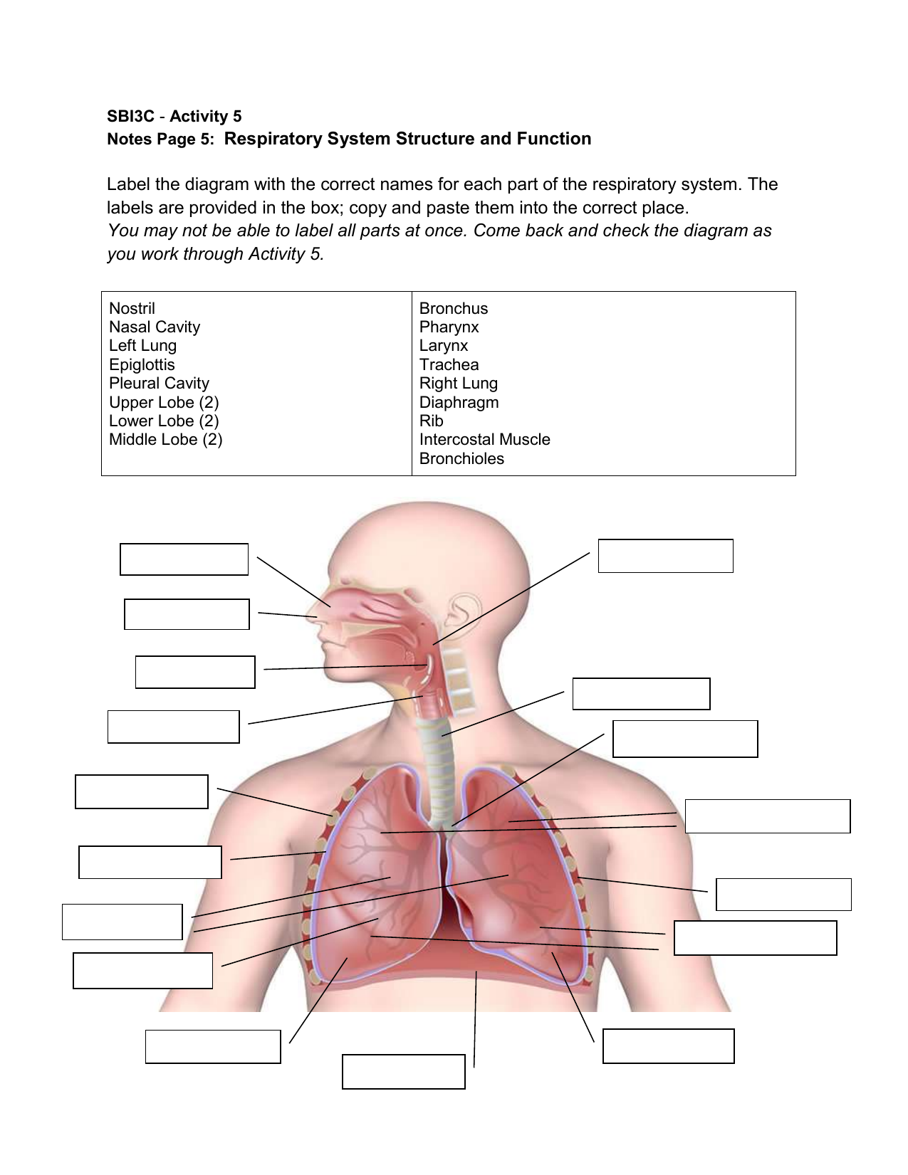
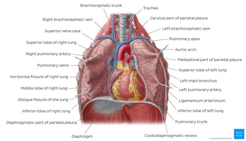
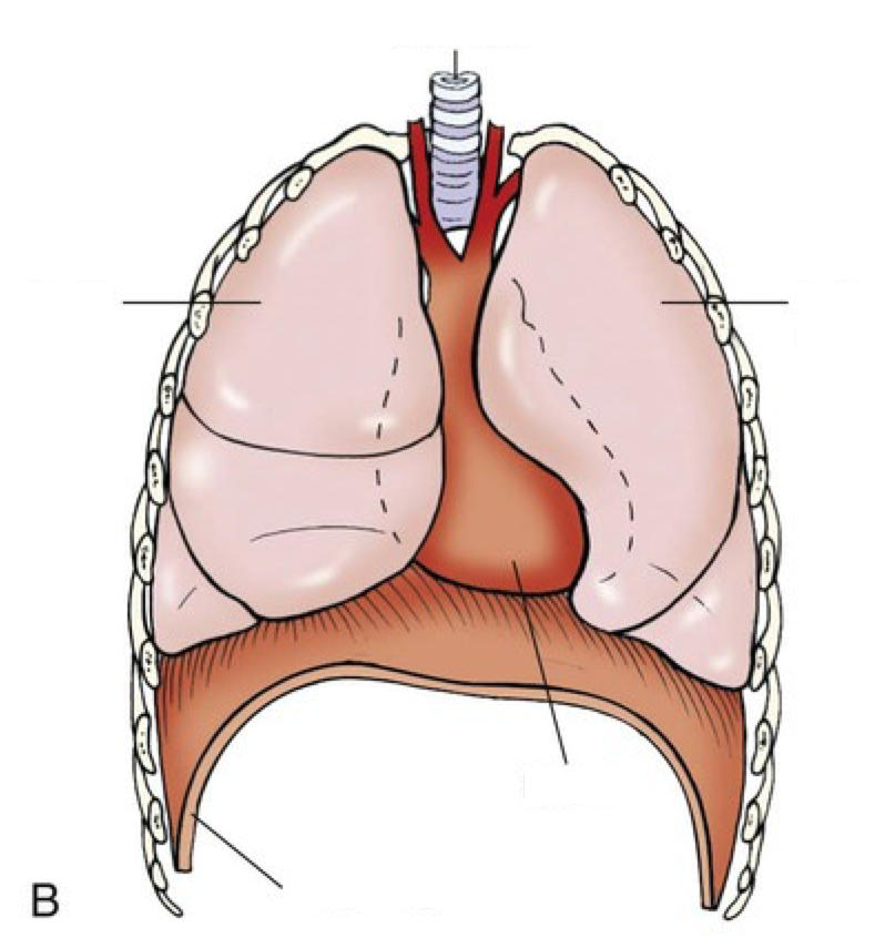
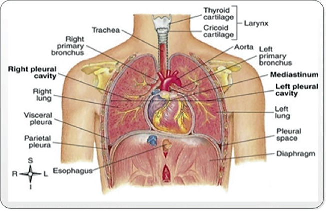
42 label the structures of the thoracic cavity.
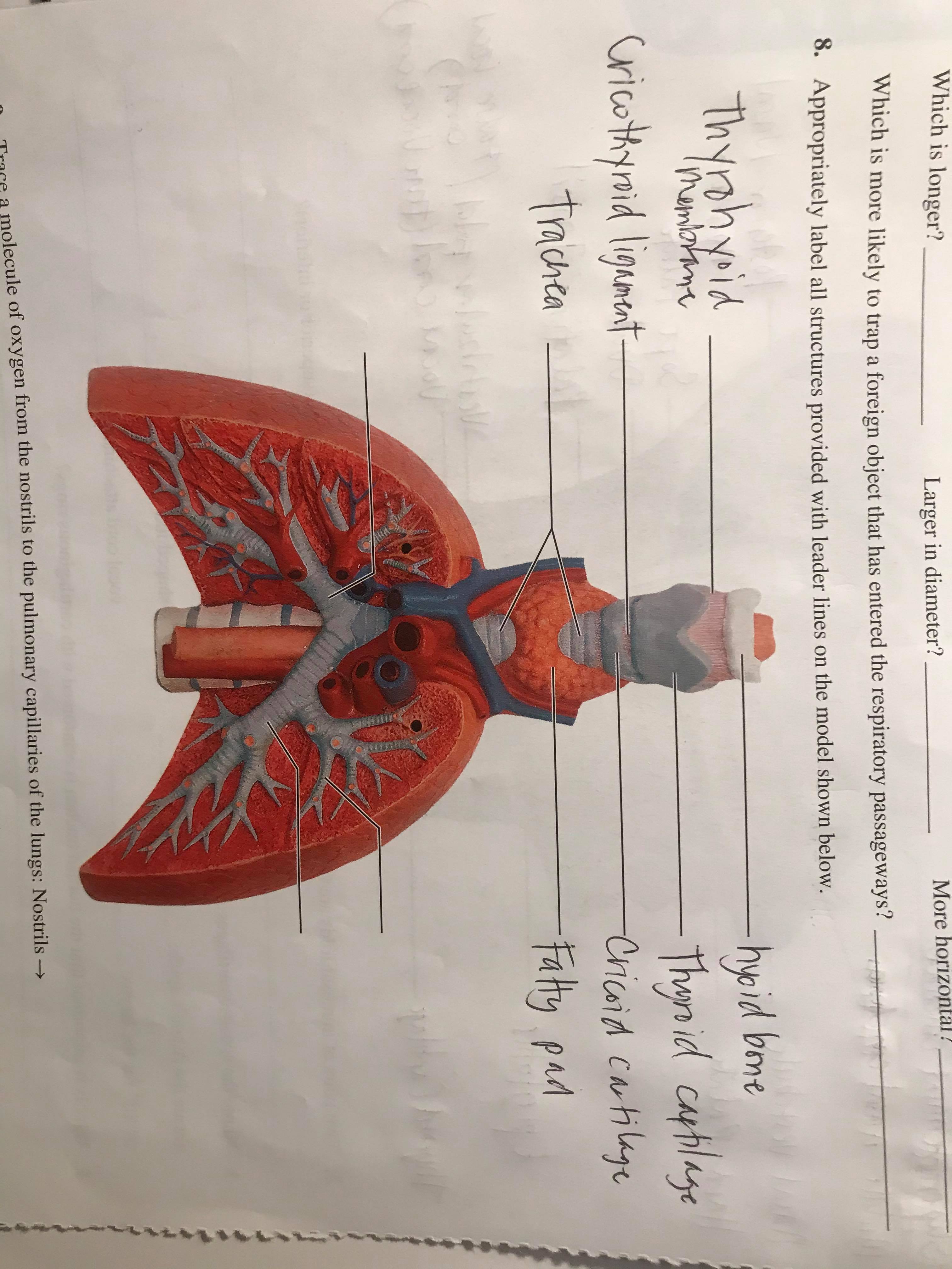
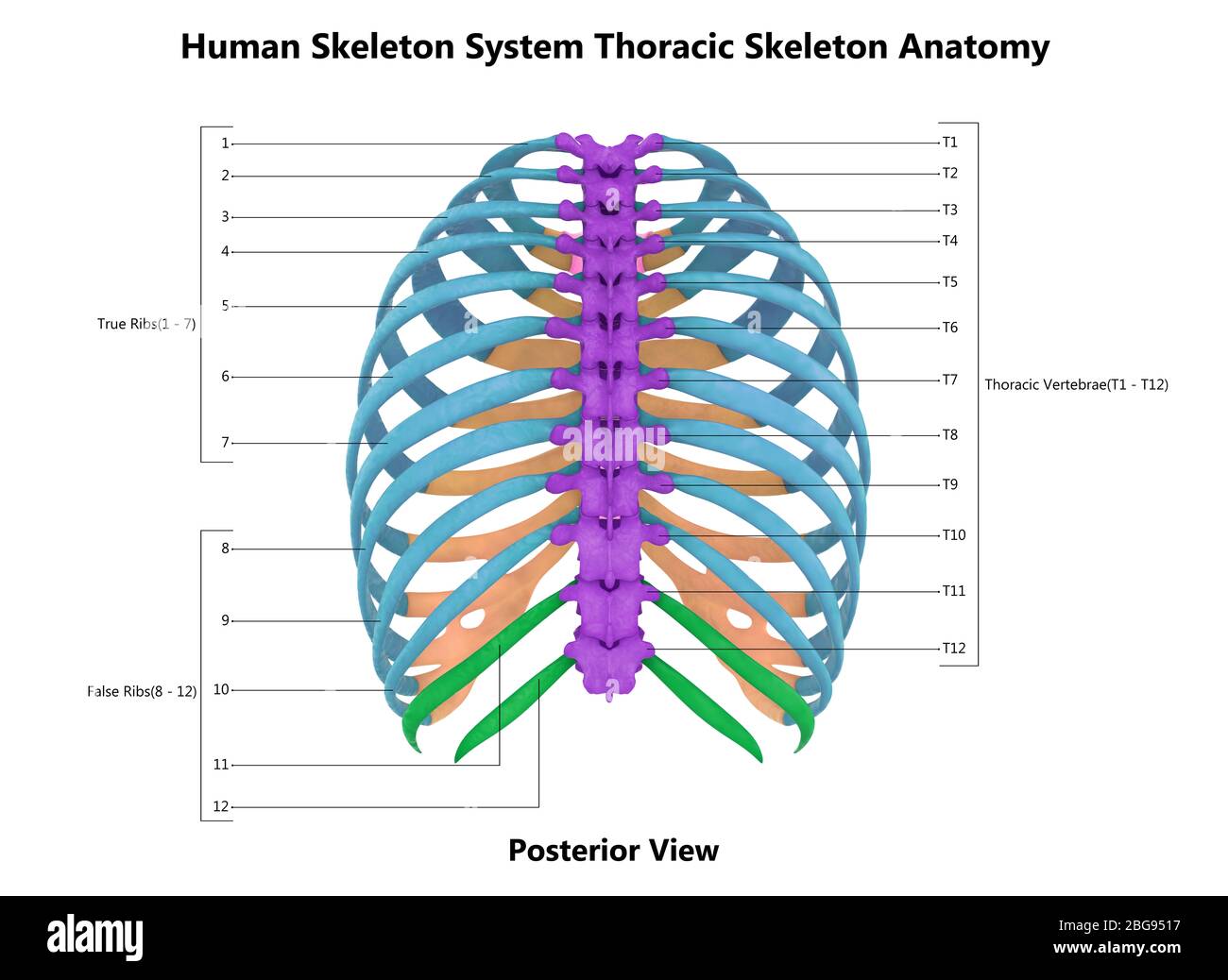
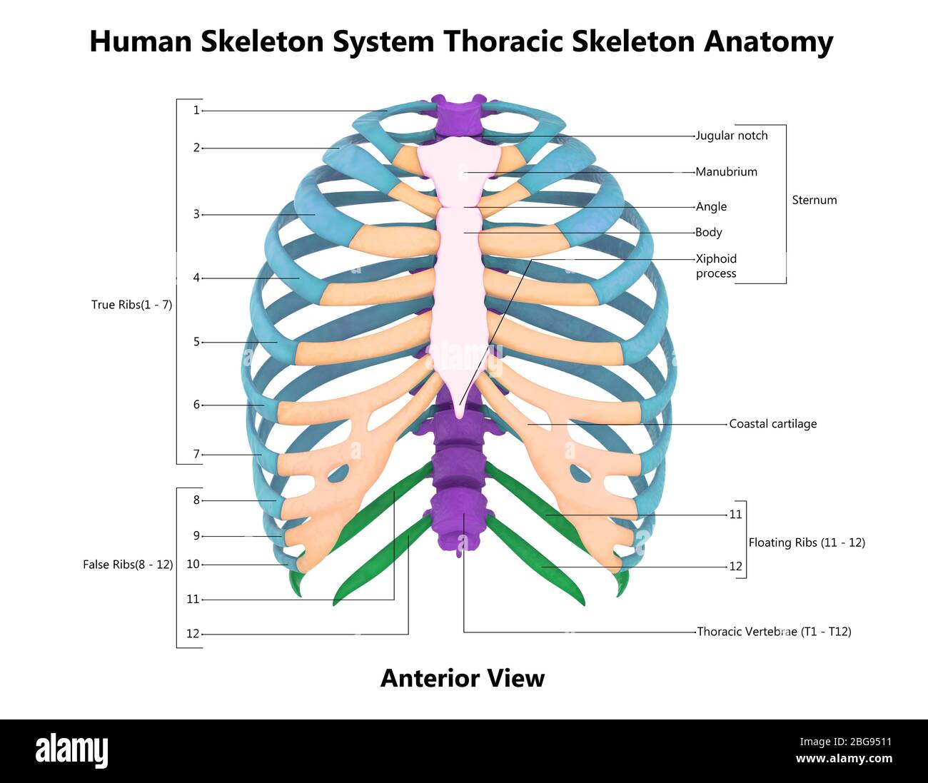
(Get Answer) - Label The Structures Of The Thoracic Cavity. Trachea ... main structures and desoribe the of the upper u and lower respieahory tract Desoribe the anatomy of the lungs and their placement within the thoracke cavity 1. Complete the following paragraph using the words peovided in the word bank:... diagram of the thoracic cavity Rat Dissection-- Thoracic Cavity & Circulatory System - YouTube . rat dissection thoracic cavity system circulatory. Thoracic Cage Labeling Quiz . cage rib diagram unlabeled anatomy bones worksheet human thoracic labeling anterior quiz bone worksheets game worksheeto purposegames via
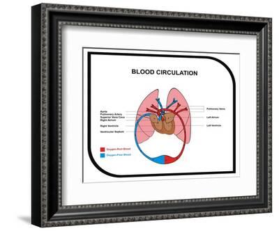
Ch. 19 Circulatory System- heart Flashcards | Quizlet The first and last structures are given. Right atrium 1. tricuspid valve 2. right ventricle 3. pulmonary valve 4. pulmonary trunk 5. pulmonary artery 6. lungs 7. pulmonary vein 8. left atrium Left atrioventricular valve
Label the structures of the thoracic cavity.
Thoracic Cavity - Introduction, Structure, Organs, and FAQs In the centre of the chest between the lungs is the mediastinum that comprises the organs that are located inside it. Structures within the thoracic cavity include: Oesophagus of the digestive system Thymus gland Vagus nerve and parasympathetic veins. Diaphragm, trachea, bronchi, lungs. The heart The superior and inferior vena cava. Body Cavities and Membranes: Labeled Diagram, Definitions First, the cranium/skull is on the outside which encloses the cranial cavity. Below the skull is 3 layers of membrane called the meninges. The 3 meningeal layers are labeled with the stars. The outermost layer of the meninges is the dura mater, which is located beneath the skull. thoracic cavity | Description, Anatomy, & Physiology | Britannica thoracic cavity, also called chest cavity, the second largest hollow space of the body. It is enclosed by the ribs, the vertebral column, and the sternum, or breastbone, and is separated from the abdominal cavity (the body's largest hollow space) by a muscular and membranous partition, the diaphragm.
Label the structures of the thoracic cavity.. Thoracic Cavity - Anatomy | Organs | Functions | 8 Types of Cavities Thoracic Cavity Right and left serous membrane cavities (contain right and left lungs) Mediastinum: Higher portion stuffed with blood vessels, trachea, esophagus, and thymus. The lower portion contains pericardial space (the heart is found at intervals the serosa cavity) 3. Serous Membranes Line of body cavities and canopy organs. Consist of Solved Correctly label the following anatomical features of | Chegg.com Biology questions and answers. Correctly label the following anatomical features of the thoracic cavity. (Not all words will be used.) Left lung Apex of heart Parietal pleura Fibrous pericardium Superior vena cava Interior vena cava Aorta Pulmonary trunk Base of heart Correctly label the following external anatomy of the anterior heart. Solved Award: 0.76 points Label the structures of the - Chegg Science. Anatomy and Physiology. Anatomy and Physiology questions and answers. Award: 0.76 points Label the structures of the thoracic cavity. Parietal pleura Visceral pleura Pleural cavity Parietal pericardium Visceral pericardium Pericardial cavity Reset Zoom. Question: Award: 0.76 points Label the structures of the thoracic cavity. Thoracic Cavity - Definition & Organs of Chest Cavity | Biology Dictionary The thoracic cavity is actually composed of three spaces each lined with mesothelium, a special film-like tissue that separates vital organs. The pleural cavities surround the lungs, while the pericardial cavity surrounds and protects the heart. These tissues in the thoracic cavity can be seen in the image below.
The Thoracic Cavity Flashcards | Quizlet precardial cavity is. a small chamber that surrounds the heart. what separates the abdominopelvic cavity from the thoracic cavity. the diaphragm. define viscera. internal organs that are enclosed by cavities. define serous membranes. serous membranes cover viscera (internal organs) and line cavities. visceral layer. thoracic cavity of a human Thoracic Cavity | Anatomy | Pinterest | Thoracic Cavity And Human Body . cavity thoracic. Circulatory System - Anatomy Of Felis Domestica anatomyofacat.weebly.com. dissection cat vessels heart system respiratory veins arteries anatomy aorta vena cava auricle male circulatory reproductive artery thoracic cats vein Label The Structures Of The Thoracic Cavity - Royal Pitch The thoracic cavity is located deep in the thorax, and is made up of several parts. The thoracic cage is comprised of four major parts: the heart, lungs, and a pericardial membrane. These areas are protected by the thoracic wall. The thorax is home to the heart, the most important organ of the thorax. thoracic cavity diagram HB Anatomy Thorax . thorax. Serous Membranes Of Thoracic Cavity . thoracic membranes serous cavity cavities label quiz flashcards. Thorax Intercostal Space . intercostal thoracic thorax drainage lymphatic ant posterior arteries thoracis azygos. Normal Chest And Abdomen Organs Medical Illustration
2 (Chap 19) Help Save & Exit Subm Check my work - chegg.com Anatomy and Physiology questions and answers. ent # 2 (Chap 19) Help Save & Exit Subm Check my work Correctly label the following anatomical features of the thoracic cavity. Base of heart Superior vena cava 3:05 Pulmonary trunk Apex of heart Aorta Inferior vena cava inces Left lung Pericardial sac Parietal pleura. (Get Answer) - Label the structures of the thoracic cavity.. Label the ... Label the structures of the thoracic cavity. a table-tennis ball is thrown at stationary bowling ball makes one-dimensional elastic collision and bounce back along the same line. compared with the bowling ball after the collision, does the table have a) larger magnitude of momentum and more... › science › refChapter One: Introduction - California State University ... cavity, the major vessels near the heart, nerves, and the esophagus. Below the thoracic cavity is the abdominopelviccavity, which contains the upperabdominalcavity,housing the digestive organs, and the inferiorpelviccavity, which holds the uterus and rectum in females or just the rectum in males. Label the specific andmajorcavities of the Unit 1 Lab Homework Flashcards - Quizlet Terms in this set (13) Label the regions of the body. Label the structures of the thoracic cavity. Label the directional terms based on the arrows. Label the body planes. Label the directional terms based on the directions of the arrows. _____ is towards the front of the body. _____ is farther from the trunk or origin of a structure.
achieverstudent.comAchiever Student: The best way to upload files is by using the “additional materials” box. Drop all the files you want your writer to use in processing your order.
› Thoracic_ExaminationThoracic Examination - Physiopedia The examiners observe the patient’s thoracic spine region and assess for the presence of deviation from normal including the thoracic spine curvatures in the frontal and sagittal planes. The overall impression of inter-rater reliability for postural observation of kyphosis and label either excessive, normal or decrease range from moderate to ...
› types › soft-tissue-sarcomaChildhood Soft Tissue Sarcoma Treatment (PDQ®)–Health ... Weiss AR, Chen YL, Scharschmidt TJ, et al.: Pathological response in children and adults with large unresected intermediate-grade or high-grade soft tissue sarcoma receiving preoperative chemoradiotherapy with or without pazopanib (ARST1321): a multicentre, randomised, open-label, phase 2 trial. Lancet Oncol 21 (8): 1110-1122, 2020.
sciencetrends.com › anatomical-body-planesAnatomical Body Planes | Science Trends Jan 01, 2019 · The cavities of the body include the dorsal cavity, the cranial cavity, the ventral cavity, the vertebral cavity, the thoracic cavity, and the abdominopelvic cavity. The dorsal cavity is one long continuous cavity that houses portions of the central nervous system including the spinal cord and brain. It is found on the body’s dorsal side.
› science › articleANATOMY AND PHYSIOLOGY - ScienceDirect Jan 01, 2005 · The thoracic cavity contains the lungs and the mediastinum, which contains the heart and its attached blood vessels, the trachea, the esophagus, and all other organs in this region except for the lungs. The abdominopelvic cavity is divided by an imaginary line into the abdominal and pelvic cavities.
Page: The Annals of Thoracic Surgery Jun 30, 2021 · The mission of The Annals of Thoracic Surgery is to promote scholarship in cardiothoracic surgery patient care, clinical practice, research, education, and policy. As the official journal of two of the largest American associations in its specialty, this leading monthly enjoys outstanding editorial leadership and maintains rigorous selection ...
Label the thoracic cavities.docx - Label the cavities below; Answer the ... Label the cavities below; Answer the questions below. Save your responses and submit in the dropbox. Understanding the Thoracic cavity, its linings and structures. In the figure above - locate the thoracic cavity. Label the structure that separates the thoracic cavity from the abdominopelvic cavity Notice the 4 colors of the thoracic cavity.
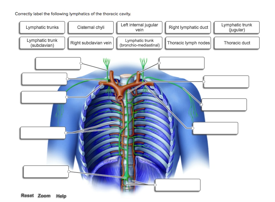
AP 2 (part 3) Flashcards | Quizlet Correctly label the following lymphatics of the thoracic cavity. ... Label the structures of the spleen. Match the lymphatic trunk with the major body region that it drains. Intestinal trunks: Drain most abdominal structures Lumbar trunks: Drain lower limbs and pelvic organs
Anatomy Chapter 1: Labeling Thoracic Cavity Diagram - Quizlet The cavities surrounding each lung parietal pleura The aspect of the pleura that does not touch the surface of the lung visceral pleura The aspect of the pleura that covers the external surface of the lung The thoracic cavity can be subdivided into... 1. mediastinum 2. left and right pleural cavities 3. pericardial cavity
Thorax: Anatomy, wall, cavity, organs & neurovasculature | Kenhub Thoracic wall The first step in understanding thorax anatomy is to find out its boundaries. The thoracic, or chest wall, consists of a skeletal framework, fascia, muscles, and neurovasculature - all connected together to form a strong and protective yet flexible cage.. The thorax has two major openings: the superior thoracic aperture found superiorly and the inferior thoracic aperture ...
Thoracic Cavity: Definition, Structure, Functions and Diseases The thoracic cavity, also known as the chest cavity, is a cavity enclosed by the ribs, the vertebral column, and the sternum or the breastbone. A muscular and membranous partition, the diaphragm, separates the thoracic cavity from the abdominal cavity.
Organs in the Thoracic Cavity - Bodytomy The ribs, vertebral column, muscles, connective tissues, and the sternum (breast bone) enclose this cavity. The thoracic cavity is lined by a serous membrane that exudes a thin fluid (serum). The chest membrane, also known as parietal pleura, extends further to cover the lungs. This part of the membrane is known as the visceral pleura.
thoracic cavity | Description, Anatomy, & Physiology | Britannica thoracic cavity, also called chest cavity, the second largest hollow space of the body. It is enclosed by the ribs, the vertebral column, and the sternum, or breastbone, and is separated from the abdominal cavity (the body's largest hollow space) by a muscular and membranous partition, the diaphragm.
Body Cavities and Membranes: Labeled Diagram, Definitions First, the cranium/skull is on the outside which encloses the cranial cavity. Below the skull is 3 layers of membrane called the meninges. The 3 meningeal layers are labeled with the stars. The outermost layer of the meninges is the dura mater, which is located beneath the skull.
Thoracic Cavity - Introduction, Structure, Organs, and FAQs In the centre of the chest between the lungs is the mediastinum that comprises the organs that are located inside it. Structures within the thoracic cavity include: Oesophagus of the digestive system Thymus gland Vagus nerve and parasympathetic veins. Diaphragm, trachea, bronchi, lungs. The heart The superior and inferior vena cava.

:watermark(/images/watermark_only_sm.png,0,0,0):watermark(/images/logo_url_sm.png,-10,-10,0):format(jpeg)/images/anatomy_term/pleura-parietalis/6qV6NXs3CWJoIPldlGAiCQ_Pleura_parietalis_01.png)












:watermark(/images/watermark_only_sm.png,0,0,0):watermark(/images/logo_url_sm.png,-10,-10,0):format(jpeg)/images/anatomy_term/diaphragma/bW4d3jaVr3PYLDmhgxbeA_Diaphragma_01.png)

Post a Comment for "42 label the structures of the thoracic cavity."