42 label eye diagram
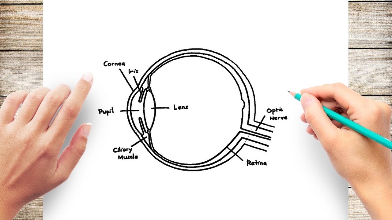
eye labeling Diagram | Quizlet delicate membrane lining the inside of the eyelids and covering the eyeball cornea fibrous transparent layer of clear tissue like a dome that covers the anterior portion of the eyeball (the iris and pupil). It is the first structure to refract (bend) light that enters the eye. sclera Tough white out covering of the eyeball choroid BYJUS BYJUS
Label the Eye Quiz - PurposeGames.com This is an online quiz called Label the Eye. There is a printable worksheet available for download here so you can take the quiz with pen and paper. From the quiz author. ... Definition And Term Match Up Game For The Eye 12p Image Quiz. Ear 11p Multiple-Choice. Label a Neuron 10p Image Quiz. Label The Neuromuscular Junction 5p Image Quiz.
Label eye diagram
Label the microscope — Science Learning Hub 08.06.2018 · All microscopes share features in common. In this interactive, you can label the different parts of a microscope. Use this with the Microscope parts activity to help students identify and label the main parts of a microscope and then describe their functions.. Drag and drop the text labels onto the microscope diagram. If you want to redo an answer, click on the box and … Anatomy of the eye: Quizzes and diagrams | Kenhub Take a look at the diagram of the eyeball above. Here you can see all of the main structures in this area. Spend some time reviewing the name and location of each one, then try to label the eye yourself - without peeking! - using the eye diagram (blank) below. Unlabeled diagram of the eye. Click below to download our free unlabeled diagram of ... Eye Diagram With Labels and detailed description - BYJUS A brief description of the eye along with a well-labelled diagram is given below for reference. Well-Labelled Diagram of Eye The anterior chamber of the eye is the space between the cornea and the iris and is filled with a lubricating fluid, aqueous humour. The vascular layer of the eye, known as the choroid contains the connective tissue.
Label eye diagram. Labeled Eye Diagram | Science Trends The iris is divided into six different layers: the anterior layer, the stroma of iris, the iris sphincter muscle, the iris dilator muscles, the anterior pigment epithelium, and the posterior pigment epithelium. The two muscles found in the eye are what control the dilating and contraction of the pupil. Labeled Eye Diagram | Human eye diagram, Eye anatomy, Diagram of the eye Heart Structure Diagram. The LS of kidney shows outer dark zone called the cortex and inner pale red zone called medulla which forms the main mass of the kidney. The medulla is made of number of pyramidal structures containing renal tubules or Nephrons projecting into the cavity towards the inner region of kidney called pelvis.This is the ... PDF Eye Diagram Labeled Game Free Books All Access to Eye Diagram Labeled Game PDF. Free Download Eye Diagram Labeled Game PDF or Read Eye Diagram Labeled Game PDF on The Most Popular Online PDFLAB. Only Register an Account to DownloadEye Diagram Labeled Game PDF. Online PDF Related to Eye Diagram Labeled Game. Get Access Eye Diagram Labeled GamePDF and Download Eye PDF Parts of the Eye Eye Diagram Handout Author: National Eye Health Education Program of the National Eye Institute, National Institutes of Health Subject: Handout illustrating parts of the eye Keywords: parts of the eye, eye diagram, vitreous gel, iris, cornea, pupil, lens, optic nerve, macula, retina Created Date: 12/16/2011 12:39:09 PM
Labeled Eye Diagram | Eye anatomy diagram, Eye anatomy, Diagram of the eye This Article is the detailed account of all the major organs that are categorized under the nine regions in the abdominal cavity 1) Stomach 2) Intestines a) Small Intestine Duodenum Jejunum Ileum b) Large Intestine Ceacum Colon (Ascending, Transverse and Descending) Rectum Anal Canal 3) Liver 4) Gall bladder 5) Pancreas 6) Spleen 7) Kidneys ... Create a Briliant Process Flow Diagram with Canva There are lots of ways to use color in a process flow diagram. You could have all the arrows in one part of the process the same color to make it clear they relate to that process. For example, you could use colors like blue and green to represent a cooling process or red and yellow to represent something being heated. To recolor any element or ... Labelled Diagram of Human Eye, Explanation and Function - VEDANTU Labeled Diagram of Human Eye . The eyes of all mammals consist of a non-image-forming photosensitive ganglion within the retina which receives light, adjusts the dimensions of the pupil, regulates the availability of melatonin hormones, and also entertains the body clock. Parts of a microscope with functions and labeled diagram 19.04.2022 · Q. Differentiate between a condenser and an Abbe condenser. Ans. Condensers are lenses that are used to collect and focus light from the illuminator into the specimen. They are found under the stage next to the diaphragm of the microscope. They play a major role in ensuring clear sharp images are produced with a high magnification of 400X and above.
Cow's Eye Dissection | Exploratorium Learn how to dissect a cow's eye in your classroom. This resource includes: a step-by-step, hints and tips, a cow eye primer, and a glossary of terms. DOC Label the Eye Diagram - Windsor C-1 School District - a thick, transparent liquid that fills the center of the eye - it is mostly water and gives the eye its form and shape (also called the vitreous humor) Answers: Label the Eye Diagram Human Anatomy. Read the definitions, then label the eye anatomy diagram below. Cornea - the clear, dome-shaped tissue covering the front of the eye. Iris How to use a swimlane diagram to improve process management 11.01.2022 · How to create a swimlane diagram in Cacoo. Creating a swimlane diagram in Cacoo, our own diagramming software, couldn’t be easier. Simply click on ‘Templates’ on the left-hand side of the page, and select either ‘swimlane flowchart’ or ‘decision flowchart.’ A readymade swimlane diagram will pop up. From there, you can edit the ... Hot Rock & Alternative Songs – Billboard The week’s most popular songs, ranked by audio and video streaming activity on leading digital music services, radio airplay audience impressions based on monitored airplay and sales data, all …
These 9 Diagrams Will Help You To Understand Makeup - ALL FOR FASHION ...
Labelling the eye — Science Learning Hub In this interactive, you can label parts of the human eye. Use your mouse or finger to hover over a box to highlight the part to be named. Drag and drop the text labels onto the boxes next to the eye diagram If you want to redo an answer, click on the box and the answer will go back to the top so you can move it to another box.

6 Best Images of Basic Parts Of Ear Worksheet - Human Ear Diagram ...
eye diagram muscle anatomy eye muscles labeled diagram eyes vision extrinsic medical alilamedicalimages illustrated. Left Eye, Lateral View schoolbag.info. anatomy eye muscles lateral left human figure facial extrinsic dynamics motion artist drawing guide classic schoolbag biology info.

Football Pitch Soccers Field Measurements Clip Art at Clker.com ...
Eye Diagram Quiz - ProProfs Try this amazing Eye Diagram Quiz quiz which has been attempted 4744 times by avid quiz takers. Also explore over 77 similar quizzes in this category. Take Quizzes. Animal; Nutrition; ... Quiz: Label The Parts Of The Eye. People say that the eyes are the windows to a person's soul. In the class today, we covered parts of the eye, and what ...

Eye and Ear Models | Eye anatomy, Anatomy models, Medical anatomy
Label the Eye Diagram | Quizlet Label the Eye Diagram | Quizlet Label the Eye STUDY Learn Write Test PLAY Match + − Created by oliviasage18 Terms in this set (8) lens ... cornea ... iris ... aqueous humor ... cilliary nerve ... pupil ... fovea ... optic nerve ...
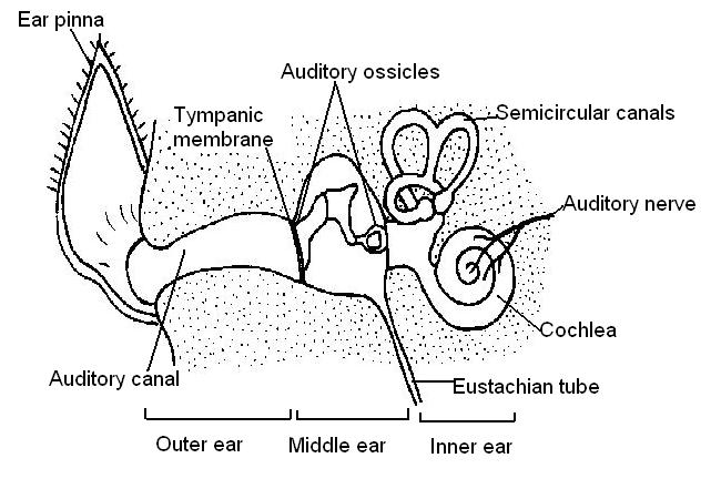
The Anatomy and Physiology of Animals/Test Yourselves/The Senses Test ...
How the Eyes Work | National Eye Institute Apr 20, 2022 · The cornea is shaped like a dome and bends light to help the eye focus. Some of this light enters the eye through an opening called the pupil (PYOO-pul). The iris (the colored part of the eye) controls how much light the pupil lets in. Next, light passes through the lens (a clear inner part of the eye). The lens works together with the cornea ...

Label Parts of the Kidney Quiz
Correctly Label the Eye Diagram Quiz - PurposeGames.com An unregistered player played the game 1 month ago About this Quiz This is an online quiz called Correctly Label the Eye Diagram There is a printable worksheet available for download here so you can take the quiz with pen and paper. Your Skills & Rank Total 0 Get started! 0

Human Eye | Eye anatomy, Basic anatomy and physiology, Biology diagrams
Eye Diagram: Label Quiz - PurposeGames.com 11. You need to get 100% to score the 11 points available. 0 favs. Add to Playlist. Game Statistics. Give a nod to the game author. Tongue Diagram: Label 4p. Ear Diagram: Label 9p.
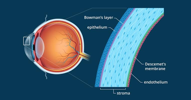
Cornea - Best Eye Care Hospital In Nagpur | Madhav Netralaya
Label Eye Printout - EnchantedLearning.com Label the Eye Diagram. Human Anatomy. Read the definitions, then label the eye anatomy diagram below. Cornea - the clear, dome-shaped tissue covering the front of the eye. Iris - the colored part of the eye - it controls the amount of light that enters the eye by changing the size of the pupil. Lens - a crystalline structure located just behind ...
How to Draw Human Eye Diagram Easy Step - YouTube
Simbrinza 1%/0.2% Eye Drops - NPS MedicineWise These dosing instructions will be printed on the label your pharmacist adds. How to use it. It is important to use Simbrinza exactly as your doctor or pharmacist has told you. If you use the drops less often than prescribed, they may not work as well and the eye problem may not improve. Using the drops more often than prescribed may not improve the eye problem any …
Post a Comment for "42 label eye diagram"